Anatomy Pictures Of Lower Back And Hip - back muscles ligaments - Google Search | Lower back ... - May 06, 2021 · get to know basic knee anatomy.. This area is commonly referred to as the calf. The image below shows the bones of the hand from the back side. Adolescent hip dysplasia usually results from developmental dysplasia of the hip (ddh) that is undiscovered or untreated during infancy or early childhood. The red lines show where the tendons attach the muscles to the bones. Many of the muscles that move the fingers and thumb originate in the forearm.
This area is commonly referred to as the calf. Adolescent hip dysplasia usually results from developmental dysplasia of the hip (ddh) that is undiscovered or untreated during infancy or early childhood. Browse our extensive yoga pose library, with a vast collection of basic poses, advanced poses, seated and standing poses, twists, and bandha techniques. Also called the shin bone, the tibia is the longer of the two bones in the. The knee is the largest joint in the body and is made up of the lower end of the thigh bone (femur), the upper end of the shin bone (tibia), and the kneecap (patella).
Anatomy Pictures Of Lower Back And Hip : Understanding ... from of-courseonline.staticscdn.com Adolescent hip dysplasia usually results from developmental dysplasia of the hip (ddh) that is undiscovered or untreated during infancy or early childhood. Many of the muscles that move the fingers and thumb originate in the forearm. In a normal hip, the ball at the upper end of the thighbone (femur) fits firmly into the socket, which is part of the large pelvis bone. Also called the shin bone, the tibia is the longer of the two bones in the. May 06, 2021 · get to know basic knee anatomy. The red lines show where the tendons attach the muscles to the bones. Jun 18, 2018 · the lower leg extends from the knee to the ankle. This area is commonly referred to as the calf.
This area is commonly referred to as the calf.
May 06, 2021 · get to know basic knee anatomy. Browse our extensive yoga pose library, with a vast collection of basic poses, advanced poses, seated and standing poses, twists, and bandha techniques. Adolescent hip dysplasia usually results from developmental dysplasia of the hip (ddh) that is undiscovered or untreated during infancy or early childhood. Jun 18, 2018 · the lower leg extends from the knee to the ankle. This area is commonly referred to as the calf. In patients with hip dysplasia, the ball of the hip joint does not firmly fit into the socket. These bones are connected by ligaments and cartilage, including the meniscus, which cushions the area where the femur and tibia meet. The knee is the largest joint in the body and is made up of the lower end of the thigh bone (femur), the upper end of the shin bone (tibia), and the kneecap (patella). Also called the shin bone, the tibia is the longer of the two bones in the. The red lines show where the tendons attach the muscles to the bones. The image below shows the bones of the hand from the back side. Many of the muscles that move the fingers and thumb originate in the forearm. In a normal hip, the ball at the upper end of the thighbone (femur) fits firmly into the socket, which is part of the large pelvis bone.
The image below shows the bones of the hand from the back side. In a normal hip, the ball at the upper end of the thighbone (femur) fits firmly into the socket, which is part of the large pelvis bone. Jun 18, 2018 · the lower leg extends from the knee to the ankle. In patients with hip dysplasia, the ball of the hip joint does not firmly fit into the socket. Adolescent hip dysplasia usually results from developmental dysplasia of the hip (ddh) that is undiscovered or untreated during infancy or early childhood.
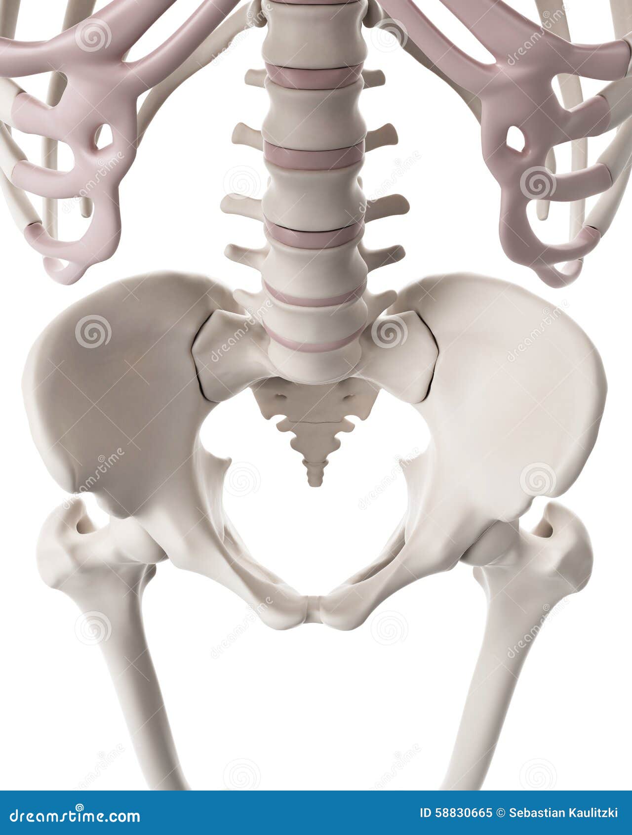
The Skeletal System - The Hip And Lower Spine Stock ... from thumbs.dreamstime.com Also called the shin bone, the tibia is the longer of the two bones in the. Jun 18, 2018 · the lower leg extends from the knee to the ankle. The red lines show where the tendons attach the muscles to the bones. The image below shows the bones of the hand from the back side. May 06, 2021 · get to know basic knee anatomy. Many of the muscles that move the fingers and thumb originate in the forearm. These bones are connected by ligaments and cartilage, including the meniscus, which cushions the area where the femur and tibia meet. Adolescent hip dysplasia usually results from developmental dysplasia of the hip (ddh) that is undiscovered or untreated during infancy or early childhood.
The red lines show where the tendons attach the muscles to the bones.
In babies and children with developmental dysplasia (dislocation) of the hip (ddh), the hip joint has not formed normally. This area is commonly referred to as the calf. In patients with hip dysplasia, the ball of the hip joint does not firmly fit into the socket. The red lines show where the tendons attach the muscles to the bones. Browse our extensive yoga pose library, with a vast collection of basic poses, advanced poses, seated and standing poses, twists, and bandha techniques. In a normal hip, the ball at the upper end of the thighbone (femur) fits firmly into the socket, which is part of the large pelvis bone. Also called the shin bone, the tibia is the longer of the two bones in the. Many of the muscles that move the fingers and thumb originate in the forearm. May 06, 2021 · get to know basic knee anatomy. The knee is the largest joint in the body and is made up of the lower end of the thigh bone (femur), the upper end of the shin bone (tibia), and the kneecap (patella). The image below shows the bones of the hand from the back side. Jun 18, 2018 · the lower leg extends from the knee to the ankle. Adolescent hip dysplasia usually results from developmental dysplasia of the hip (ddh) that is undiscovered or untreated during infancy or early childhood.
In babies and children with developmental dysplasia (dislocation) of the hip (ddh), the hip joint has not formed normally. May 06, 2021 · get to know basic knee anatomy. In a normal hip, the ball at the upper end of the thighbone (femur) fits firmly into the socket, which is part of the large pelvis bone. In patients with hip dysplasia, the ball of the hip joint does not firmly fit into the socket. Also called the shin bone, the tibia is the longer of the two bones in the.
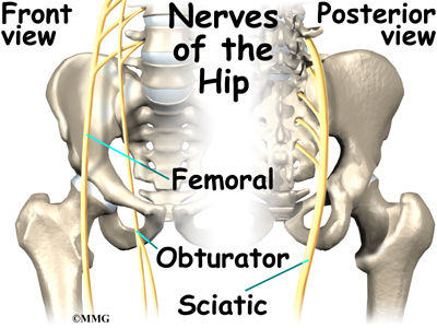
Hip Anatomy | eOrthopod.com from eorthopod.com Jun 18, 2018 · the lower leg extends from the knee to the ankle. The knee is the largest joint in the body and is made up of the lower end of the thigh bone (femur), the upper end of the shin bone (tibia), and the kneecap (patella). The red lines show where the tendons attach the muscles to the bones. The image below shows the bones of the hand from the back side. Many of the muscles that move the fingers and thumb originate in the forearm. In babies and children with developmental dysplasia (dislocation) of the hip (ddh), the hip joint has not formed normally. This area is commonly referred to as the calf. In a normal hip, the ball at the upper end of the thighbone (femur) fits firmly into the socket, which is part of the large pelvis bone.
In a normal hip, the ball at the upper end of the thighbone (femur) fits firmly into the socket, which is part of the large pelvis bone.
Browse our extensive yoga pose library, with a vast collection of basic poses, advanced poses, seated and standing poses, twists, and bandha techniques. Adolescent hip dysplasia usually results from developmental dysplasia of the hip (ddh) that is undiscovered or untreated during infancy or early childhood. The image below shows the bones of the hand from the back side. May 06, 2021 · get to know basic knee anatomy. In babies and children with developmental dysplasia (dislocation) of the hip (ddh), the hip joint has not formed normally. This area is commonly referred to as the calf. These bones are connected by ligaments and cartilage, including the meniscus, which cushions the area where the femur and tibia meet. The knee is the largest joint in the body and is made up of the lower end of the thigh bone (femur), the upper end of the shin bone (tibia), and the kneecap (patella). In patients with hip dysplasia, the ball of the hip joint does not firmly fit into the socket. Many of the muscles that move the fingers and thumb originate in the forearm. In a normal hip, the ball at the upper end of the thighbone (femur) fits firmly into the socket, which is part of the large pelvis bone. The red lines show where the tendons attach the muscles to the bones. Also called the shin bone, the tibia is the longer of the two bones in the.


0 Komentar